Highlights of our Work
2024 | 2023 | 2022 | 2021 | 2020 | 2019 | 2018 | 2017 | 2016 | 2015 | 2014 | 2013 | 2012 | 2011 | 2010 | 2009 | 2008 | 2007 | 2006 | 2005 | 2004 | 2003 | 2002 | 2001
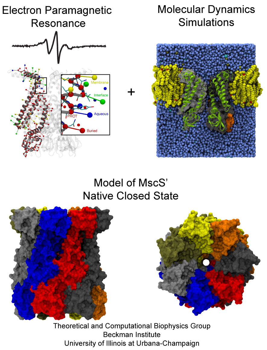
image size:
227.2KB
made with VMD
The environment of cells can undergo drastic changes, for example
from dry to wet, in which case cells shrivel or swell. However,
they are protected from bursting by a system of safety valves in
their cellular membranes that open and release cellular content.
Some of the valves open already at low membrane tension, but only
little, others open only at higher tension, but wide and without
filtering outflow. The mechanosensitive channel of small
conductance, MscS, is a low pressure safety valve in bacterial cells
(see the Feb
2007 highlight, "Observing and Modeling a Crucial Membrane Channel", the May 2006
highlight, "Electrical Safety Valve", and the Nov 2004
highlight, "Japanese Lantern Protein"). MscS is able
to rescue cells about to burst by releasing small solutes through a
large and transient opening in the cell membrane, thereby relieving
internal pressure. The only way to learn how MscS performs this vital
task is by determining its atomic-level structure under native
conditions. However, the only structure available for MscS was
obtained for the purified and crystallized protein; inspection of
the structure left doubt that it shows a functional protein, i.e., a
closed safety valve. Now a team of experimentalists and modelers
report
the structure of MscS seen in its natural membrane
environment. In their approach, simulations incorporate information
from so-called paramagnetic resonance measurements experiments. This
finding is yet another case where the combination of modeling and
observation offers entirely new close-up views of living cells (more
on our MscS website).


