Highlights of our Work
2024 | 2023 | 2022 | 2021 | 2020 | 2019 | 2018 | 2017 | 2016 | 2015 | 2014 | 2013 | 2012 | 2011 | 2010 | 2009 | 2008 | 2007 | 2006 | 2005 | 2004 | 2003 | 2002 | 2001
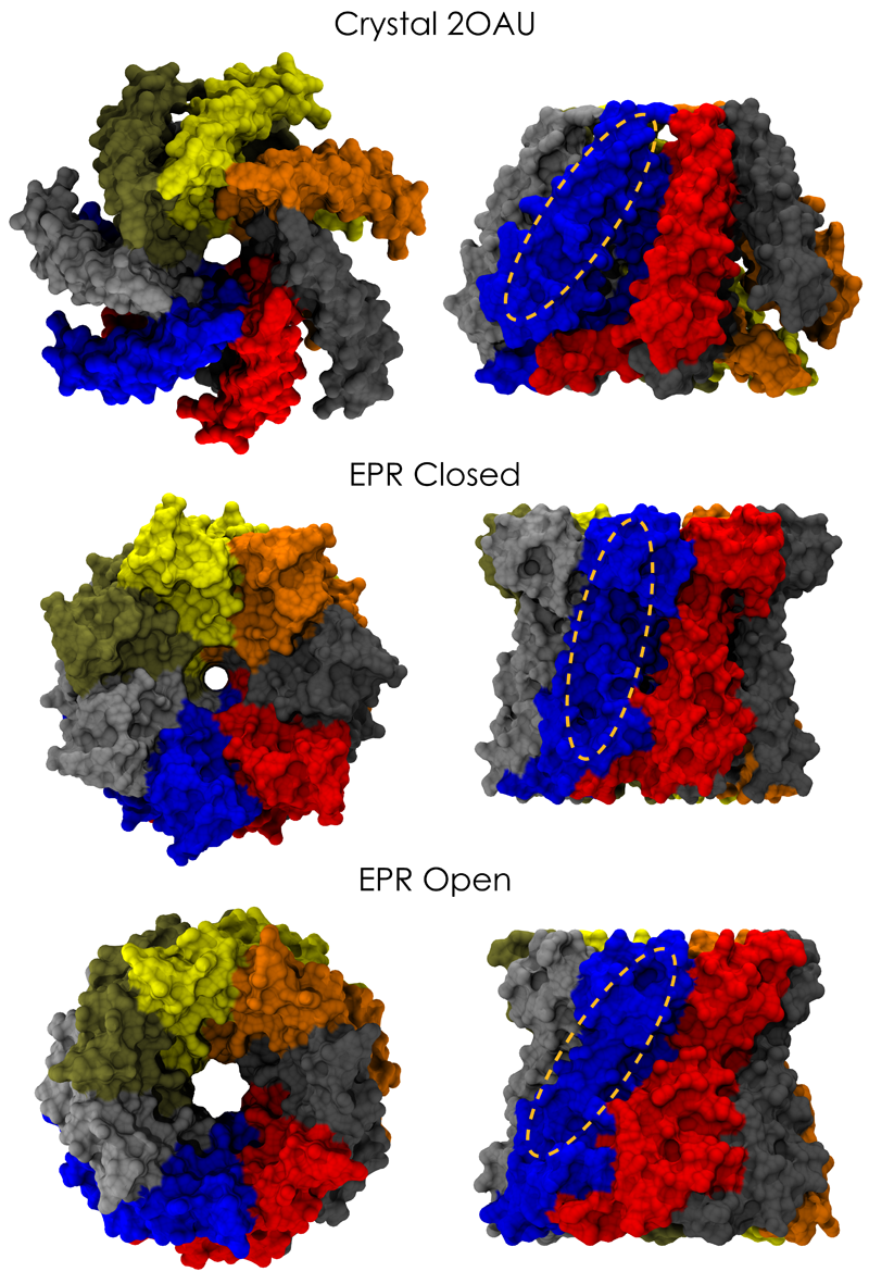
image size:
661.9KB
made with VMD
Biological cells are surrounded by a highly versatile, yet very feeble cellular membrane and need to balance
differences between the cell's interior and exterior that otherwise would burst the membrane. For example, the osmotic
pressures inside and outside the cell need to be closely balanced. Thousands of proteins in the membrane act as gatekeepers,
opening pores that can also act as safety valves, helping to reduce the interior-exterior difference in pressure rapidly. One
such protein, the mechanosensitive channel of small conductance MscS (see the Mar 2008 highlight, "Observation and
Simulation Depict Cell's Safety Valve", the Feb
2007 highlight, "Observing and Modeling a Crucial Membrane Channel", the May 2006
highlight, "Electrical Safety Valve", and the Nov 2004
highlight, "Japanese Lantern Protein") opens in response to cellular membrane tension
generated due to a drastic imbalance in osmotic pressure as it arises when a bacterial cell suddenly finds itself in fresh
water, rather than a highly saline physiological medium. The MscS channel widens then to jettison molecules out of the
cell and reduce tension on the cellular membrane quickly. In a recent report, researchers have combined experimental data
from electron
paramagnetic measurements and computer modeling to reveal in atomic detail how MscS opens and closes its channel. Combining
measurement and modeling, the researchers established a highly resolving computational microscope, unmatched by existing
microscopes (more on our MscS website).


