Highlights of our Work
2026 | 2025 | 2024 | 2023 | 2022 | 2021 | 2020 | 2019 | 2018 | 2017 | 2016 | 2015 | 2014 | 2013 | 2012 | 2011 | 2010 | 2009 | 2008 | 2007 | 2006 | 2005 | 2004 | 2003 | 2002 | 2001
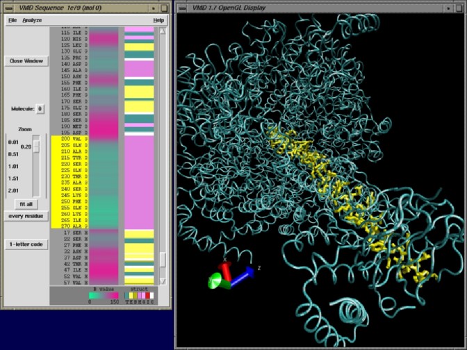
image size:
105.4KB
made with VMD
Sequence and structure are complementary properties of proteins that are critical for our understanding of the molecular machinery of living cells. VMD now makes the study of both easier than ever by integrating its structure viewing capabilities with a new sequence viewing window. Protein residues selected in the structure window can be highlighted in the new sequence window; conversely, the three-dimensional structure of segments selected from the new sequence window can be displayed in the structure window. With its powerful molecular graphics capabilities and its existing and planned sequence viewing tools, VMD has become an essential tool in biomedicine.
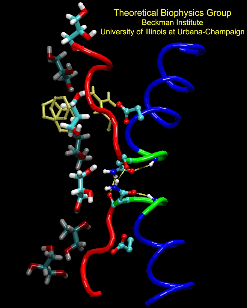
image size:
246.0KB
made with VMD
Aquaporins are channel proteins abundantly present in all life forms, for example, bacteria, plants, and in the kidneys, the eyes, and the brain of humans. These proteins conduct water and small molecules, but no ions, across the cell walls. Their defective forms are known to cause diseases, e.g., diabetes insipidus, or cataracts. The molecular modeling program, NAMD, along with large parallel computers at the Pittsburgh and Illinois supercomputing centers, permitted researchers now to model aquaporins in the natural environment of membrane and water in one of the largest molecular dynamics simulations ever (over 100,000 atoms). The simulations revealed in unprecedented detail how cells conduct water and glycerol, a molecule that serves cells' metabolism. The simulations provided a movie of the entire conduction process.
image size:
78.8KB
made with VMD
Mechanical force is seen today as a key component of molecular processes in cells: forces can be signals as in touch receptors, products as in muscle action, and substrates as in the matrix surrounding moving cells . This view of molecular processes is the result of a series of ground-breaking investigations that have become possible only recently and is rapidly turning into a new field, mechanobiology (see recent review). A collaboration with V. Vogel and coworker (U. Washington, Seattle) has investigated structural changes accompanying stretch-induced unfolding events in type III fibronectin, a protein of the extracellular matrix, explaining the design of these proteins.
image size:
155.3KB
made with VMD
Most of energy consumed by living species on earth stems ultimately from the sunlight harvested by photosynthetic cells. The light harvesting apparatus works like a solar cell, and optimizing its efficiency for the various habitats of life forms was a key strategy for survival of the fittest. After exploring for decades the role of individual molecules in light harvesting, chlorophylls, carotenoids, and small aggregates of these, researchers in the Theoretical Biophysics Group have now been able to put their findings together to calculate the overall efficiency and operational characteristics of a particular light harvesting system, that of purple bacteria. Based on structural biology, spectroscopy, and quantum mechanical calculations, a kinetic scheme was devised to show how evolution, like a good engineer, optimized the overall function of the system. The results are described in a recent paper, as well as in a review article .
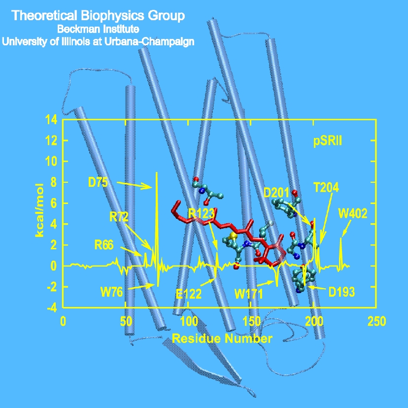
image size: 331.7KB
Perception of light and color permits humans to enjoy art, though the sense evolved more likely to better find ripe fruits. The recognition, centuries ago, that the wondrous sense of color comes from three visual receptors to which is added in the eye a fourth black & white one is one of the major achievements in the history of science. The visual receptors of all animals rely on one molecule for light absorption, retinal. How do the receptors tune then their spectral sensitivity? Exploiting a similarity of visual receptors to proteins in an archaebacterium, Natronobacterium pharaonis, researchers have finally been given an opportunity to answer this question quantitatively. In the bacterium, two structurally almost identical proteins absorb maximally light of 497 nm and 568 nm wavelength. X-ray crystallography and advanced quantum chemical studies could explain the difference and pinpoint to the protein side groups (see figure) that actually tune the spectra.
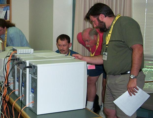
image size: 41.0KB
Our low-cost cluster of 32 Athlon PCs (see February 2001 highlight) has been in constant use by local users, providing a substantial and very cost-effective boost to our group's large-scale simulation capabilities. To satisfy demand, we have added two additional 32-processor clusters with higher performance at an even lower cost. On this platform, the freely available simulation code NAMD can complete a 1 nanosecond simulation of the 60,000 atom aquaporin-1 water channel with full electrostatics and constant pressure in a single week. We have given three tutorials, both filled to capacity, introducing participants to cluster hardware and software with the aid of a hands-on session assembling and installing four-processor clusters (see photo).
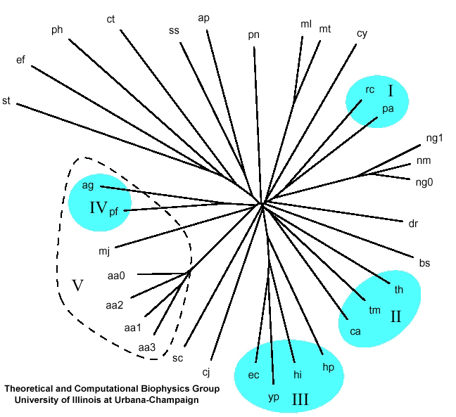
image size: 172.1KB
Organisms evolve according to the Darwinian principle by optimizing their traits through mutations of their genomes. In the past this principle had been rather narrowly applied to single genes and the associated single proteins. Knowledge of the sequences of the entire genomes of organisms encourages today a broader perspective: evolution should improve the entire context in which proteins function in organisms, for example, entire metabolic or signaling pathways. A new method describing the tree of evolution of organisms includes accordingly entire groups of genes that consolidate a cellular reaction network. The new phylogenetic analysis of metabolic networks has been applied in a recent publication to electron-transfer and amino acid biosynthesis networks yielding a more comprehensive understanding of similarities and differences between organisms.
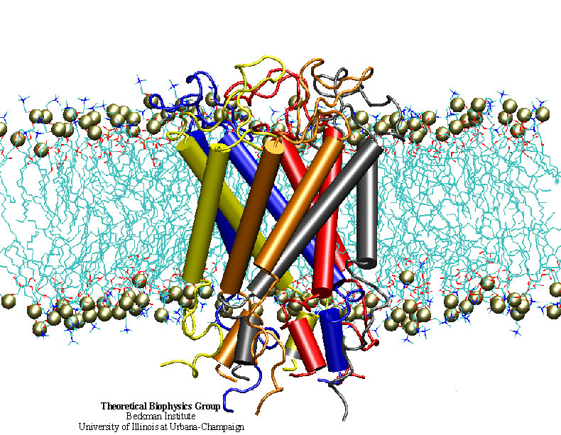
image size:
247.9KB
made with VMD
"How do you feel?" Biologists now have an answer that may surprise you. Our sense of touch relies upon the fact that cells in our fingertips can sense the pressure from a tabletop and transmit a signal to the brain. But until recently, the molecular mechanism for turning the stretching of a cell membrane into a cellular signal was unknown. An important step in understanding this process was the discovery of a protein known as a the mechanosensitive channel of large conductance, or MscL. Though this protein has been studied primarily in bacteria, homologues exist in all major kingdoms of life. Researchers in the Theoretical Biophysics Group have used molecular dynamics simulations to study, at the atomic level, how MscL opens in response to pressure changes. Models of MscL will give us new insight, not only into how we feel, but also how our hearts beat and how we keep our balance. Feel better now? ( more, publication )
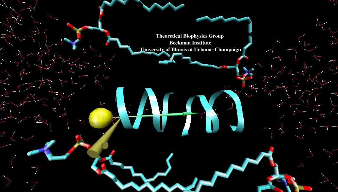
image size:
173.4KB
made with VMD
"If I could just get my hands on that protein!" Single molecule manipulation techniques like atomic force microscopy have brought us closer to this frequently expressed wish. These techniques, however, do not "see" the atomic level detail needed to relate mechanism to protein architectures. True, computational methods do illuminate the elusive protein structures, but are limited to static structures, or trajectories yielded by weeks-long simulations. Now, with the advent of inexpensive, high-performance computing, interactive manipulation of molecular dynamics simulations has become a reality. Linking advanced molecular graphics with ongoing molecular dynamics simulations, and utilizing a haptic device to connect forces from a user's hand with forces in the simulation, researchers can interact with "live" proteins. The new methodology is described in a recent publication and the figure shown here demonstrates a Cl- ion being pulled through a gramicidin A channel (see a 3.8mb Streaming Video).

click for an image gallery
Our UIUC College of Engineering 2001 Open House event featured a simulation of the regulation of genetic expression in bacteria using NAMD and VMD. We showed a model of a repressor protein that shuts down an active DNA site by forcing the DNA to adopt a looped form. The combination of a detailed all-atom model for the protein with a coarse-grained model for the DNA loop helps us to reduce the computational time required to simulate the behavior of this protein-DNA system. With a second project we illustrated how through Interactive Molecular Dynamics (IMD) one can manipulate manually, with force feedback, simulated biomolecules for the purpose of drug design. IMD, via a haptic input device, relies on researchers' sense of touch in their quest to understand how biomolecules work. Finally, we presented BioCoRE, a collaborative research environment for structural biology within which researchers can visualize information, share resources and interact with each other and with structural biology tools via a common infrastructure and across time zones and continents.
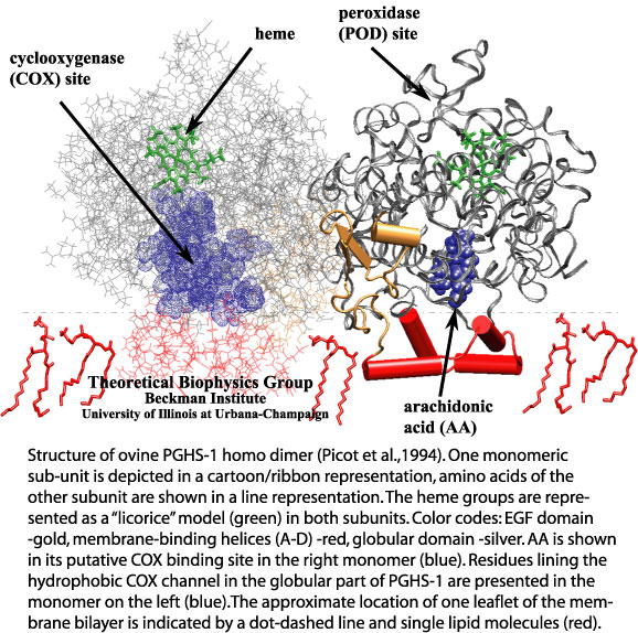
image size:
128.8KB
made with VMD
Aspirin, the widely used pain killer, has revealed many beneficial effects such that it has attracted renewed attention. It has become known that aspirin acts as an inhibitor to prostaglandin synthase. Pharmacological researchers have succeeded to improve aspirin's effect by synthesizing analogue compounds, so-called superaspirins, that target the right type of prostaglandin synthase in the body. The continuing effort has been supported by basic research on the properties of prostaglandin synthases. Molecular dynamics simulations, carried out with our molecular dynamics program NAMD, have investigated how prostaglandin synthases select their substrates, arachidonic acid, through a binding channel that acts as a filter for compounds with the right stereochemical properties. The figure, taken from a recent publication, and made with our graphics program VMD, shows one monomeric subunit (in a cartoon/ribbon representation) of the ovine PGHS-1 homo dimer. To see both subunits click on the image. [More Information]
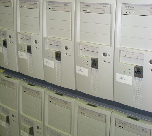
image size: 25.8KB
We have installed a low-cost cluster of 32 PCs with 1.1GHz Athlon processors, 256MB of RAM, and switched fast ethernet. On this new platform, the freely available simulation code NAMD runs 1 ns of our 92K atom ApoA1 PME benchmark in 8 days with 70% efficiency, the equivalent of a 100 processor Cray T3E. The new machine will be useful for simulations such as the stretching of the muscle protein titin. This work seeks to examine in atomic detail the dynamics and structure-function relationships of this 30,000 amino acid long filament in muscle contraction and elasticity. The cluster also provides a powerful engine for interactive simulations. The Linux-based Scyld Beowulf operating system (see Scyld's press release regarding NAMD) makes the entire cluster appear to computational biologists as a single machine.
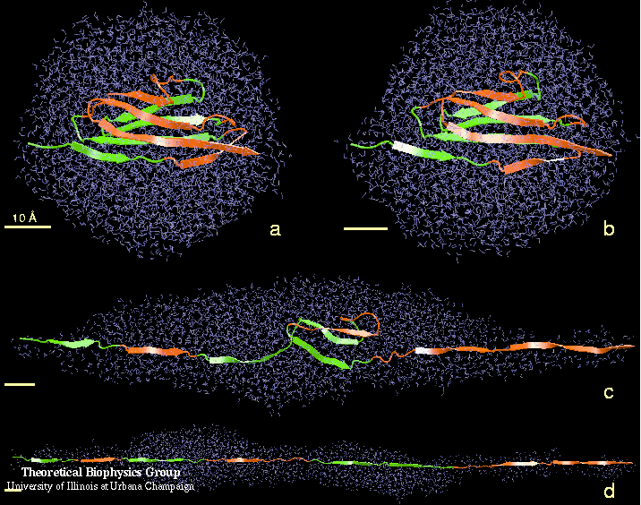
image size:
106.6KB
made with VMD
Water can act as a conformational lubricant for protein folding. The giant muscle protein titin is a roughly 30,000 amino acid long filament which plays a number of important roles in contraction and elasticity. For example, upon stretching in muscle some of titin's protein domains can unfold one-by-one permitting titin to retain elastic properties in muscle over a very wide range of length. To examine in atomic detail the dynamics and structure-function relationships of this behavior, SMD simulations of force-induced titin domain unfolding were performed in close collaboration with atomic force microscopy observations. The simulations led to the discovery that water molecules play an essential role in breaking sets of hydrogen bonds that control the unfolding of titin's domains (see resulting publication).
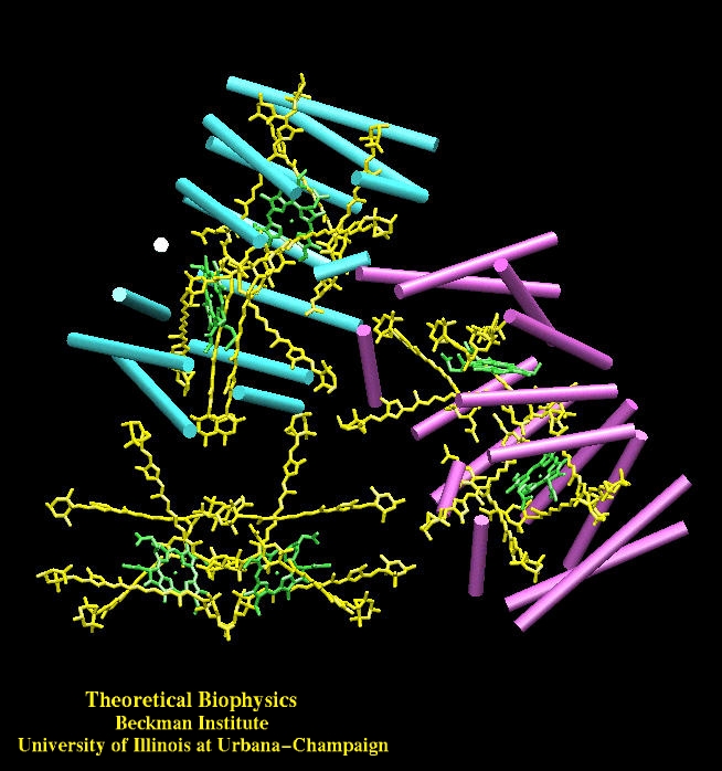
image size:
195.0KB
made with VMD
A photosynthetic variant explains how nature harvests sunlight. Most photosynthetic life forms employ mainly chlorophylls for absorbing sun light and fueling its energy into their metabolism. However, dynoflagellates, e.g., the species Amphidinium carterae, utilizes predominantly carotenoids for light absorption. Biophysicists have computed the electronic properties of a key light harvesting protein of Amphidinium carterae, peridinin-chlorophyll-protein, and in comparing their results with properties known for more conventional photosynthetic systems have learnt how biological evolution molded efficient solar batteries and what principles guided the evolution of photosynthesis (see resulting publication).
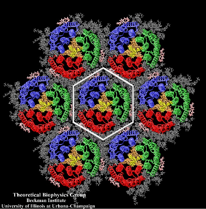
image size:
319.7KB
made with VMD
Halobacteria and people have in common a type of membrane protein (retinal proteins) that contains the molecule retinal and is activated by sun light. In Halobacteria the proteins serve the bacterium's energy needs as well as steer the bacterium into areas that are lit appropriately. In people, other vertebrates and also invertebrates, the protein provides the light receptors for vision. In Halobacteria one retinal protein, bacteriorhodopsin, forms a two-dimensional crystal in the bacterial cell wall. Relying on observations from crystallography and electron microscopy, computer modeling as [reported = purple membrane] recently could reconstruct the entire purple membrane, combining protein, lipids, and water. Here, for the first time, a fully functional biological system is known precisely, i.e., every atom type and position being identified. Now the mechanism underlying an integral biological function, in this case light absorption leading to the pumping of protons across a membrane, can be deduced from physical principles. More can be found on our website.


