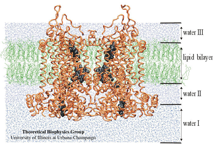
Cytochrome bc1 complex
 A higher-resolution TIFF version
(1.3M) of the above
figure, without the text and call-out lines, is available.
A higher-resolution TIFF version
(1.3M) of the above
figure, without the text and call-out lines, is available.
Description
Model of the structure of the cytochrome bc1 complex from chicken heart mitochondria with the iron-sulfur protein in the position proximal to the Qo site, inserted in the membrane bilayer. The core proteins and other domains of the bc1 complex on the matrix side of the membrane are not included. The protein backbone is represented by orange tubes; the Fe2S2 cluster and the heme groups are rendered as grey spheres; the lipid molecules are drawn with green lines, and the solvent water molecules are shown as blue lines and dots. The shading of the solvent reflects the procedure of building the model. Water molecules that were used for solvation of the lipid bilayer are shown as light blue lines (water II and III), while the water molecules added to the system to completely solvate the inter-membrane domains of the bc1 complex after placing it into the membrane are shown as darker blue dots(water I).
The above solvated structure of the bc1 complex in a phospholipid bilayer contains a total of 206,720 atoms. A subset of 91,061 atoms was actually simulated with 45,131 moving atoms.


