Movie Gallery - Ribosome
All movies and images were made using VMD. Download movie files via the movie file type (.mpg, .mov) after the movie title (right-click in Windows; control-click on Macs). To view original movie files, you may need to try the Quicktime or VLC movie players. You may wish to also visit TCBG's YouTube movie gallery to view science movies. All movies are copyrighted by TCBG.
 |
Desktop MDFF (.mpg, 29M) | ||||
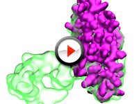
| The movie demonstrates how the once computationally-intensive Molecular Dynamics Flexible Fitting (MDFF) method can now be achieved on a desktop in a matter of minutes using the NAMD software. Depicted is an HIV inactivating protein (in purple) being morphed by MDFF towards a simulated EM density map (in green) derived from the same protein in a different state, taken from the PDB database. Taking advantage of NAMD and GPU computing, the MDFF simulation shown only took 10 minutes to finish on a desktop. The greater speed provided by NAMD stands to advance cryo-EM modeling using the MDFF method. | |||||
 |
Ribosome-Translocon-Membrane Complex (.mpg, 29M) | ||||
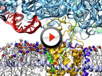
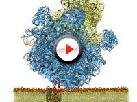
| The ribosome, protein factory for all cells, is seen in blue docked to the a protein-conducting channel, the tranlsocon, in gold. The translocon is in the process of inserting a new protein (green) into the membrane (white with red/blue/tan spheres), aided by a specific part of the ribosome in red. Membrane proteins play a key role in many human diseases, and understanding how they are inserted and then fold is critical to developing novel therapeutics targeting them. | |||||
 |
Symmetry and Functions of Enzymes (.mpg, 3.4M) | ||||
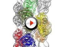
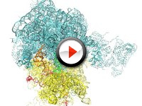
| The movie shows structures of a nitrilase multi-enzyme system forming helical fiber (nine subunits in blue, red, gray, orange, yellow, tan, silver, green and white inside an electron microscopy density map) as revealed by using the Molecular Dynamics Flexible Fitting method. Knowing the symmetry of nitrilase helps in understanding how nitrilase converts toxins into non-toxic materials for bacterial cells, a process which can inform industrial efforts to remove toxins. | |||||
 |
New Protein Exiting from a Ribosome (.mpg, 8.6M) | ||||
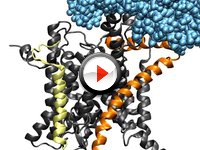
| Ribosome (in blue) makes cell proteins that then have to be moved or "translocated" by another protein, SecY (in gray, orange, yellow) across the cell membrane. The movie depicts the simulated translocation of a growing protein (in green) from the ribosomal exit tunnel into the channel of SecY. | |||||
 |
Molecular Dynamics Flexible Fitting (.mpg, 5.4M) | ||||
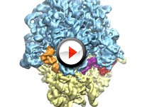
| Ribosome (in blue, gold) makes cell proteins from amino acids delivered by tRNAs (orange, green, purple) and EF-Tu (red), and is a popular target for antibiotics research. A method for capturing ribosome in action is shown; the method reveals movement of a protein (blue, curly) into the ribosome. | |||||
 |
View of Ribosome (.mpg, 5M) | ||||
| This movie shows an equilibration of the ribosome. | |||||
 |
Ribosome-SecY Complex (.mpg, 9.1M) | ||||
| The ribosome is seen bound to the protein-conducting channel SecY, which rests inside a lipid bilayer. The complex was built using cryo-EM data and MDFF. | |||||


