Movie Gallery - Channel Proteins
All movies and images were made using VMD. Download movie files via the movie file type (.mpg, .mov) after the movie title (right-click in Windows; control-click on Macs). To view original movie files, you may need to try the Quicktime or VLC movie players. You may wish to also visit TCBG's YouTube movie gallery to view science movies. All movies are copyrighted by TCBG.
 |
Water Channels in Cell Membranes (.mpg, 13.1M) | ||||
| Every second about a billion water molecules (red and white spheres) pass through a channel formed in the middle of an aquaporin protein as shown by white (nonpolar) and green (polar) areas, and charged areas in blue (positive) and red (negative). The yellow sphere highlights the path of a single water molecule. | |||||
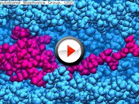 |
How Bacteria Makes a Tail (.mpg, 8M) | ||||
| Depicted is the movement of flagellin (in red) through surrounding flagellum (in blue) to the tip of the tail in bacteria. At the tip, the flagellin folds into place to construct a full tail. Bacteria then use the tail to move in search of nutrients. Understanding this process informs our knowledge of bacterial self-assembly and transport. | |||||
 |
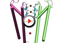
Drying Out Potassium (.mpg, 1M) | ||||
| Understanding how ions move through nanopores can aid in designing tools for sequencing DNA. The movie shows a potassium ion (blue sphere) and its water shell (red and white around blue sphere) moving through a raw pore covered with dangling oxygens (purple spheres), dangling silicons (green spheres), and non-dangling atoms (gray). The water shell is partially removed and the ion adsorbed near the surface of the pore. After some time, the ion is released and the hydration shell restored. | |||||
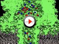 |
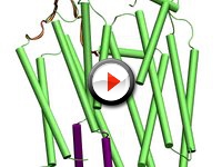
Electrical Current through a Bacterial Toxin (.mpg, 2.9M) | ||||
| Alpha-hemolysin, a protein manufactured by bacteria that forms pores in cell membranes, can be used to detect molecules because they block electrical current through the pore. The movie shows a computer simulation in which the motion of ions from salt dissolved in the water can be seen. The positively charged potassium ions (in blue) move upward through the alpha-hemolysin (in green), while the negatively charged chloride ions (in red) move downward, constituting an electric current. | |||||
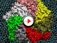 |
Alpha-Hemolysin: A Protein That Kills (.mpg, 4M) | ||||
| The bacterium Staphylococcus aureus, a common cause of infection in humans, secretes a toxin known as alpha-hemolysin that kills human cells by forming holes in their membranes, through which chemicals essential to survival leak out. The movie illustrates the structure of the hole, which is created when identical proteins released by the bacterium (in pink, yellow, gold, red, grey, green, and white) assemble in groups of seven in the cell's membrane (in cyan). | |||||
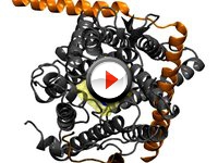 |
Translocation of a Polypeptide Helix through SecY (.mpg, 3.1M) | ||||
| Proteins serve a variety of roles in the cell. One protein, SecY, manages the movement or 'translocation' of other protein segments across the membrane (i.e., a polypeptide helix). The helix (in blue), forces open the pore ring (yellow) and displaces the plug of SecY (red), thus completing a protein translocation event. | |||||
 |
Sugar Permeation through LacY (.mpg, 2.8M) | ||||
| Lactose is induced through LacY by SMD. The kink of helix I (in pink) facilitate smooth sugar passage. Lactose undergoes a rotation passing the central binding cavity. | |||||
 |
K+ Permeation through Kv1.2 Channel (.mpg, 1.6M) | ||||
| The movie shows simulation of K+ conduction through Kv1.2 under a voltage bias. The conduction occurs via the knock-on mechanism as suggested by Hodgkin and Keynes about 50 years ago. | |||||


