Calcium Binding Proteins: Calmodulin
Calcium binding proteins regulate many important cellular processes such as smooth muscle contraction and the crossbridge motion in skeletal muscle. Calmodulin is a rather ubiquitous calcium-sensing protein belonging to a class of loop-helix-loop cation binding proteins of similar structure and function. Molecular Dynamics simulations of calmodulin uncover important dynamical aspects of the regulatory mechanisms of this class of proteins (Pascal-Ahuir, Mehler, Weinstein: Molecular Engineering 1, 231-247, 1991). For instance, the structural flexibility of the central alpha-helical tether is believed to be an essential element in the calcium-dependent recognition of target-peptides.Simulations of Calmodulin
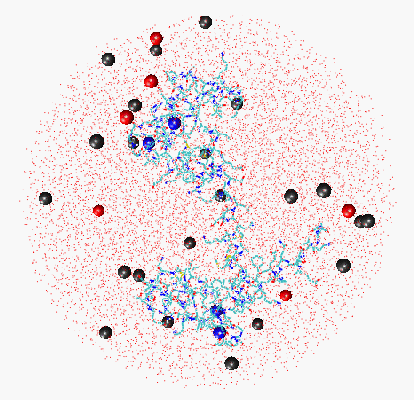 Fig. 1: Click here to get a 45.1 kByte image of calmodulin, solvated in a 44 A radius sphere of waters (only oxygens shown) with counterions at physiological ionic strength 75 mM, ~33,000 atoms (red: Cl-, grey: Na+, blue: Ca++).
In collaboration with Prof. Harel Weinstein and Prof. Ernest Mehler (Mount Sinai School of Medicine, City University of New York) we have carried out a 3 ns simulation of the system on the Cray T3D at Pittsburgh Supercomputer Center. We studied the reorientation of the two major domains of calmodulin and the flexibility of the tether. The simulations shed light on the time-dependent availability of various target-specific structures of calmodulin. The central tethering helix, which earlier has been shown to undergo large conformational changes upon binding to target proteins, bends over its length and the two calcium-binding domains reorient with respect to each other. This rearrangement of the structure brings the domains to a more favorable position for target binding, poised to achieve the orientation observed in the CaM-myosin-light-chain-kinase complex (Ikura et al., Science (1992) 256:632).
Our 3 ns trajectory provides a near-complete sampling of the counterion distribution about the protein. In Fig. 2, which made the cover of the Biophysical Journal issue, we compare the three-dimensional histogram of sodium ions sampled from the 3 ns trajectory with the electostatic potential surrounding the protein calculated from the Poisson-Boltzmann equation at the ionic strength of the simulation. The Figure demonstrates that the counterions are more localized in regions of negative electrostatic potential.
 Fig. 2: Click here to get a 118 kByte image of counterion distribution and electrostatic potential surrounding calmodulin, as shown on the cover of Biophysical Journal, April 1998.
Left: Three-dimensional histogram of sodium ion distribution in calmodulin's vicinity sampled from the 3 ns trajectory. The colors code for the ion density values 1.0 M (red) and 3.0 M (yellow). Right: Contours of constant sodium concentration and electrostatic potential near the surface of the negatively charged protein. The contour was calculated with the program Grasp at 75 mM ionic strength. The colors code for the ion density values 0.1 M (blue) and 0.2 M (green), and 0.5 M (yellow).
Reference:
Willy Wriggers, Ernest Mehler, Felicia Pitici, Harel Weinstein, and Klaus Schulten. Structure and Dynamics of Calmodulin in Solution. Biophysical Journal 1998, Vol. 74, pp. 1622-1639.
If you use diagrams or material from this site, I ask that you cite the home page and author, or the appropriate source publication in your work. Copyright 1997-1998. All rights reserved.


