Symbiont Bacteria
Bacterial communities within the human body greatly influence human health and play a significant role in diseas e predisposition, pathogenic, physical fitness, and dietary responsiveness. Importantly, bacteria utilize highly co operative macromolecular machines to accomplish many cellular functions. Here we seek to understand, with molecular and atomistic fidelity, two such machines: the cellulosome and the chemosensory array, which underlie the phenomen a of bacterial plant fiber degradation and chemotaxis respectively.
Biofuels: Bacteria can make a living off a very wide range of food sources. This agnosticism enables them to, among other things, serve as essential symbionts in animal digestive tracts where they assist their hosts in d egrading cellulose fibers into metabolizable compounds. In particular, bacteria in the rumen of the cow face an esp ecially tough job (see Tight Job in the Gut), digesting the hardy cellulose fibers of grasses. Key to their task are molecular tentacles on the cell s urface of certain gut bacteria, so-called cellulosomes (pictured right), which develop a tight grasp on cellulose a nd then effectively cleave the molecules. In general, human gut bacteria (and their role in the broader human micro biome) are one of the most intensely researched topics in medicine.
Bacterial Chemotaxis: Bacteria monitor their environments and respond by way of a fundamental sensory cap ability known as chemotaxis---one of the best studied behavioral systems in biology. Chemotactic responses in bacte ria involve large complexes of sensory proteins, known as chemosensory arrays, that process the information obtaine d from the bacteria's habitat to determine its swimming pattern. In this sense, the chemosensory array functions as a bacterial brain, transforming sensory input into motile output. Despite great strides in the understanding of ho w the chemosensory array's constituent proteins fit and work together, a high-resolution description has, until rec ently, remained elusive (see Computing the Bacterial Brain). Here we are combining computational and experimental techniques to explore in detail the molecular mechanisms underlying sensory signal transduction and amplification within this amazing biological appar atus.
Spotlight: Bacteria Swim and Tumble (Jan 2007)
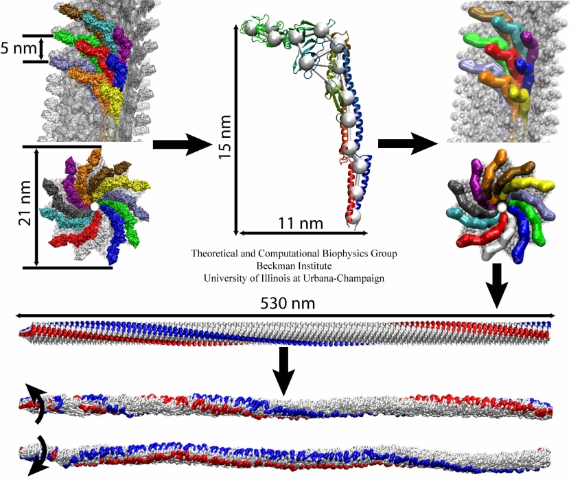
image size:
299.9KB
made with VMD
The bacterial flagellum is a large biomolecular assembly used by many types of bacteria as a helical propeller for forward swimming and turning. The flagellum is remarkable in that its properties differ greatly depending on the direction in which it is rotated, allowing the bacterium to switch between swimming straight ("running") and turning ("tumbling"). The mechanics of the flagellum are of interest both to biologists and mechanical engineers. The molecular mechanisms of the transition in the flagellum between running and tumbling modes is unknown. Because of the flagellum's size (several micrometers in length) and composition (made up of 30,000 protein subunits) it presents a challenge to computational modeling. Researchers have now achieved an advance describing the flagellum in both its running and tumbling state. For this purpose, the researchers developed a computational model of the system that glosses over atomic level detail, but resolves the shapes of all proteins making up a bacterial flagellum, simulating a simplified version of the system using the program NAMD. The results, reported recently, showed that the flagellum's transition between swimming straight and tumbling is triggered by friction due to the water around the bacterium. More information on the flagellum project can be found here.


