Graphene nanopores
DNA translocation through graphene nanopores
Nanopores have emerged as promising next generation devices for single-molecule detection, analysis and DNA sequencing. One of the drawbacks of solid-state nanopores is that the membrane thickness is about 100 times the distance between two bases in a DNA molecule. Graphene is a material with extraordinary electrical and mechanical properties. It is the thinnest known material with thickness equal to one atomic layer of carbon (0.3 nm), which is comparable to the DNA base pair stacking distance of 0.35 nm, making the graphene nanopore a promising device for DNA sequencing.
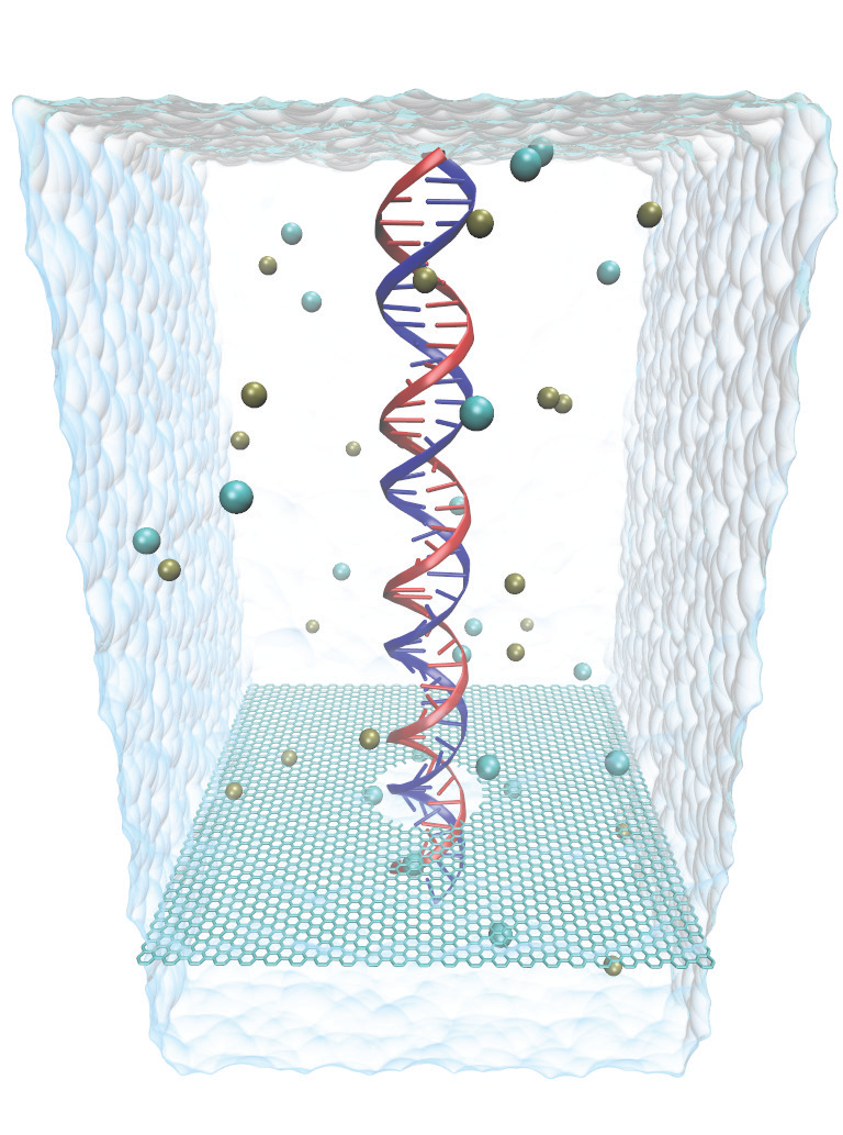
Fig. 1 - Atomic model of graphene nanopore system.
In this website, we briefly describe the kinetics of DNA translocation through graphene nanopores. For more details, take a look at the publications and links on this web site or contact the investigators listed below.
Open pore characteristics
The study of open pore characteristics of graphene membranes is perfomed to access the ability of MD simulations to faithfully reproduce electric field- driven transport of ions through nanopores. A series of all-atom MD simulations are performed to compute the resistance of the pore for pore diameters in the range of 2-7 nm. The pore resistance seems to depend inversely on the square of the pore radius, consistent with experimental observations. The potential maps (see Fig. 2) illustrate that most of the potential drop arises across the membrane and the drop becomes sharper near the membrane as the size of the pore decreases.
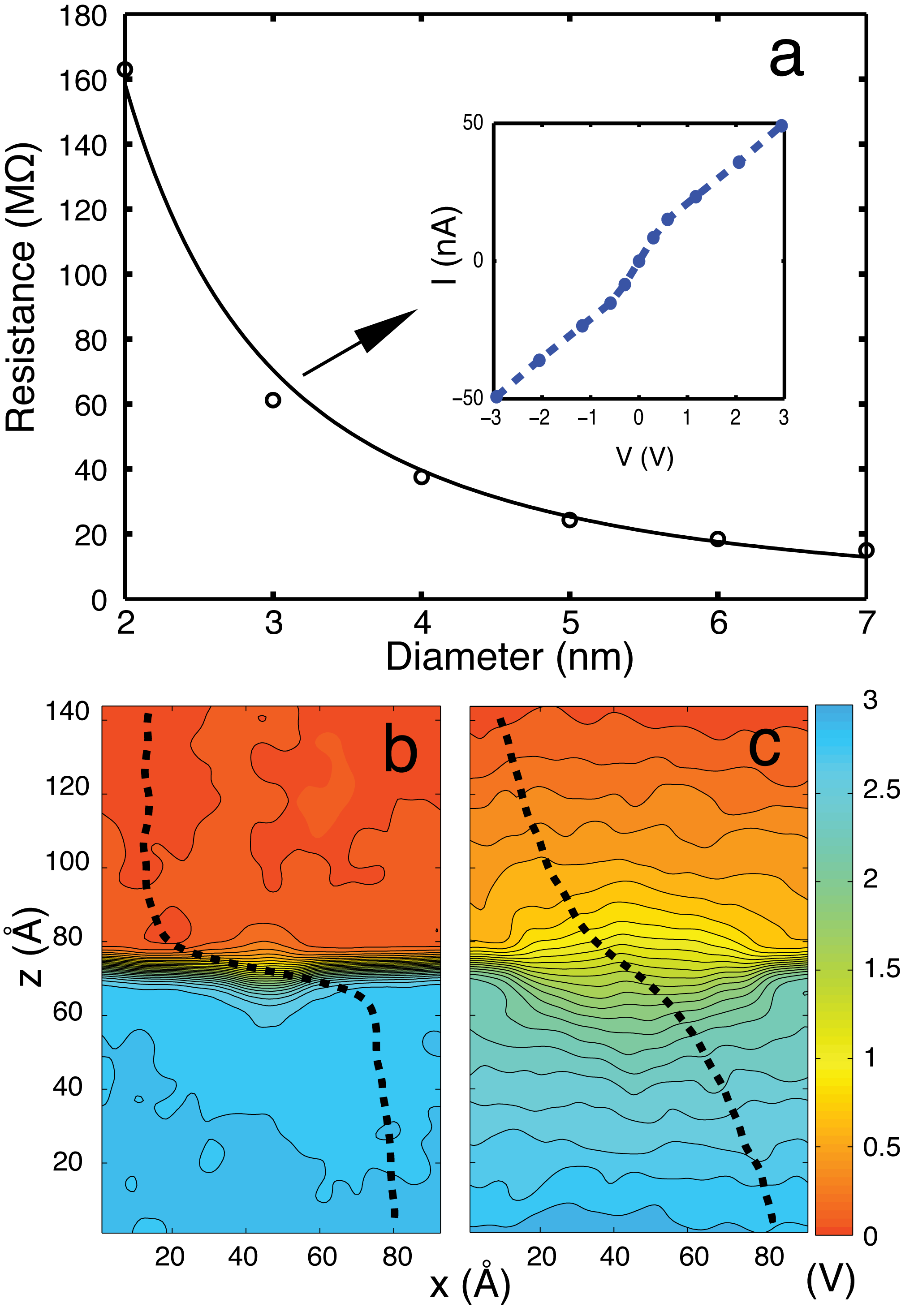
Fig. 2 - Open pore characteristics of graphene nanopores. b) and c) are the potential maps for 2 nm and 7 nm pore diameter respectively. The applied bias voltage was 3 V.
Voltage-dependent Kinetics of DNA Transport
The kinetics of dsDNA translocation was studied at bias voltages of 4.3, 2.5 and 0.8 V. DNA was placed in all simulations in a linear head-tail configuration at the pore mouth. As seen in Fig. 3, a characteristic blockade of the ion current occurs when the DNA is in the nanopore and the current returns back to the open pore value, once the DNA exits the pore. The blocking is also more effective at lower bias voltages. The movies and the different slopes in the center of mass (CoM) illustrate the strong hydrophobic interaction between DNA and graphene at low applied bias which causes the DNA to stick to the graphene membrane.
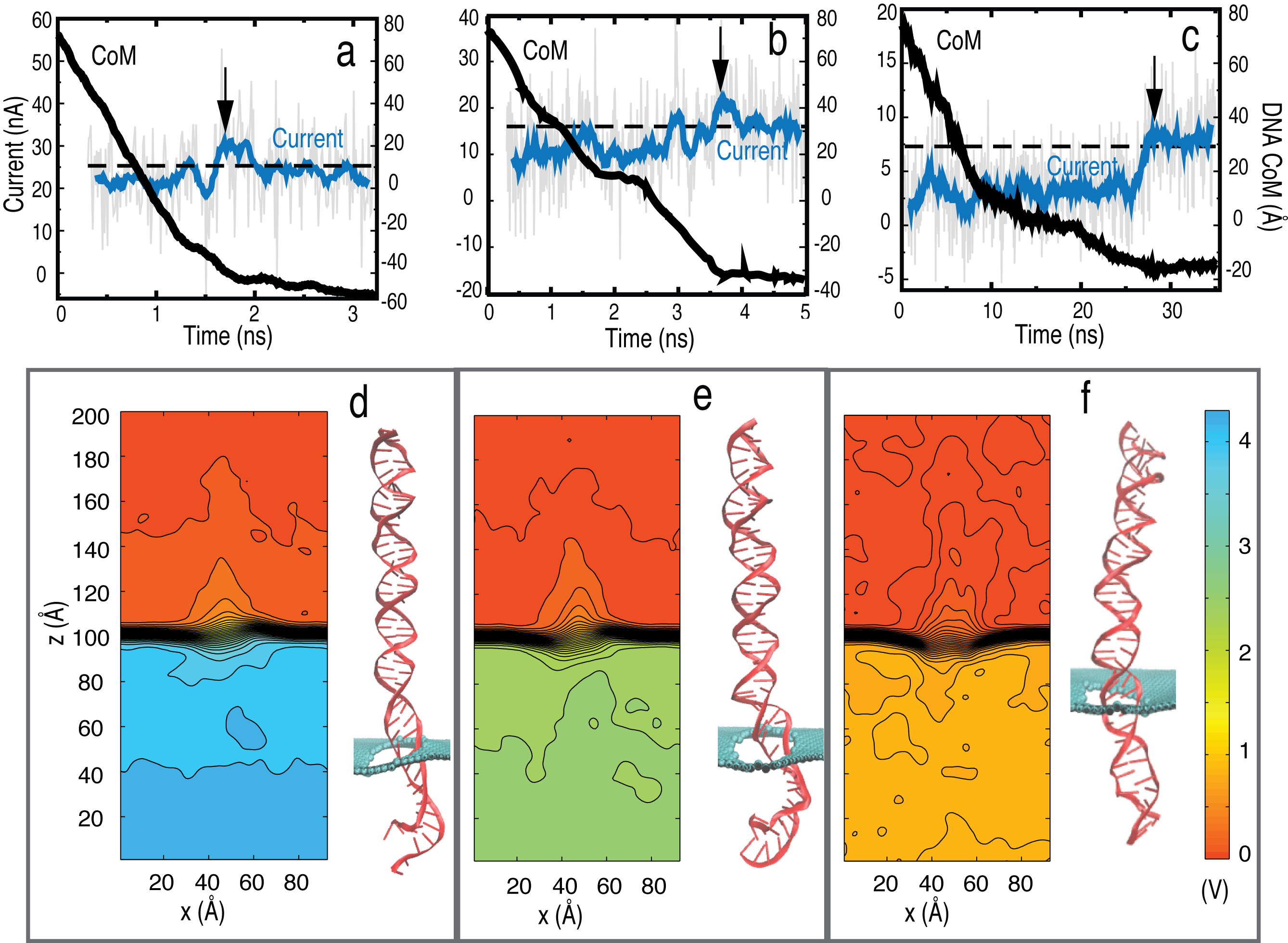
Fig. 3 - Electrophoresis of dsDNA through graphene nanopores for bias voltages
of a) 4.3 V, b) 2.5 V and c) 0.8 V. The potential maps along with a
snapshot of DNA in the pore is shown for d) 4.3 V, e) 2.5 V and f) 0.8 V.
Detecting A-T and G-C base pairs
A-T and G-C base pairs stretch to a different degree for a particular range of bias voltages. This can be attributed to the difference in the molecular structure of A-T and G-C base pairs, A-T has one less intermolecular hydrogen bond compared to G-C base pair causing A-T base pairs to stretch more readily. The ability of graphene nanopores to tailor the electric field near the pore mouth allows one to discriminate between A-T and G-C base pairs which is the first step towards sequencing DNA.
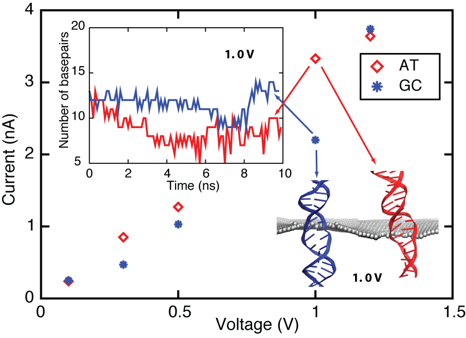
Fig. 4 - Ionic current for poly(A-T) and poly(G-C) strands.
Movies
MOVIE_1.mpg (1.3 MB): Shows the DNA translocation trajectory at an applied bias of 4.3 V.
MOVIE_2.mpg(1.6 MB): Shows the DNA translocation trajectory at an applied bias of 2.5 V.
MOVIE_3.mpg (1.6 MB): Shows the DNA translocation trajectory at an applied bias of 0.8 V.
MOVIE_4.mpg (8.3 MB): Shows the DNA translocation trajectory of poly(A-T) sequence at an applied bias of 1.0 V.
MOVIE_5.mpg (8.7 MB): Shows the DNA translocation trajectory at poly(G-C) sequence at an applied bias of 1.0 V.
Publications
Investigators
Related TCB Group Projects
Page created and maintained by Chaitanya Sathe.


