|
|
|
|
|
Kinesin |
|
|
|
Materials on this site are copyrighted!

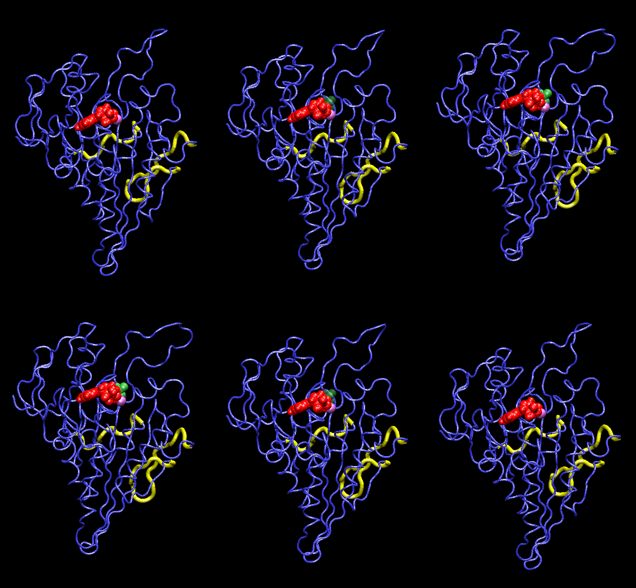
This movie (2.11 MB, gif format) shows conformational changes induced in the kinesin structure (blue) by the additional gamma phosphate (green) of ATP. The shown conformation oscillates between model structures of ADP-bound and ATP-bound kinesin which have two putative microtubule binding regions (yellow) superimposed. ADP and the associated Mg ion are shown in red and purple, respectively. The movements are visualized with the program vmd.
|
|
|
|
|
|
Materials on this site are copyrighted.
wriggers@ks.uiuc.edu