The Sequence window is used to list the residue sequences of proteins and the base sequences of nucleic acids, and to select residues/bases from the sequence list for highlighting in the 3-D structure in the main VMD window. When residues are selected in the main VMD window, the corresponding residue is highlighted in the sequence list in this window. Color-coded protein structure information is displayed for amino-acid residues, and B-factor information is displayed for all residues. In this section, ``residues'' refers both to amino acid residues in proteins, and to nucleotide bases with associated backbone in DNA and RNA molecules.
The Sequence window contains a vertical listing of the residue sequence of a loaded molecule. The Molecule pop-up menu control chooses which molecule to display the sequence of, the current 'top' molecule is displayed the first time the Sequence window is opened. The name and molecule number of the sequence displayed is shown in the title frame of the Sequence window.
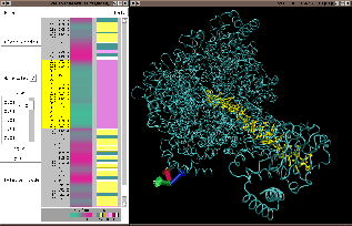
For each residue displayed, the window lists: residue number, residue name/code, and chain letter. If no chain is specified, chain letter is set to ``X''. To the right of this are two color coded columns, ``B value'' and ``struct''. ``B-value'' shows the contents of the B-value (temperature factor) field. The ``struct'' field shows protein secondary structure; select Help:Structure Codes from the window menu, or see Table 5.5, for an explanation of the single letter codes in the color key.
|
Click anywhere in the vertical listing with the left mouse button to highlight one residue. Click and drag with the left mouse button to highlight multiple residues, shift-click to add a single residue to the current selection, shift-click and drag to add multiple residues to your selection, right-click to de-select a residue. Highlights appear as thick yellow ``Bonds'' representations, these can be changed or turned off.
The Zoom slider, and the Fit all, Every Residue buttons, zoom in and out of a long sequence list to allow viewing and selecting from the entire list all at once. To represent more than 40 residues on the window, the text list seems to ``skip'' residues, but selections, highlights and color-coded data are still active for all residues.
By setting the Zoom slider to a value smaller than 1.0, or by pressing the Fit all button, more or all of the sequence information for a large molecule can be seen at once. To show a text line for every residue in the sequence (zoom factor = 1.0), click on the Every Residue button. The Zoom slider can be dragged with the left mouse button (to re-scale sequence smoothly) or it can jump to a given value by clicking along the slider track with the middle button (this is useful to work more quickly with very long sequences).
For a multi-thousand residue protein with Fit all selected, hundreds of residues can be selected at once, and trends in B-value and structure across the entire protein sequence can be detected. In the screen-shot above, a section of 70 residues with lower B-values than surrounding sequence is selected, by dragging a rectangle around the green stretch in the B-value column.
Other controls include:
To clear all highlights, reselect the current molecule from the Molecule pop-up menu. To turn the highlight representation off completely for a given molecule, find the representation in the Graphics window which the Sequence window has created (appears with ``Bonds ColorID 4'') and set the style to ``none''. To change highlighting style, set this same representation to your preferred style and coloring. The selection for this representation will still change whenever the sequence window selection changes. Example application: specify Multiple Frames in the Trajectory tab of the highlight representation. This will display the trajectory motions of the residues clicked on in the main VMD window, or in the Sequence window.