Many enzymes utilize a tripeptide called glutathione (![]() -Glu-Cys-Gly), or GSH for short. The tripeptide is unusual in that the first amino acid, glutamic acid, is linked to cysteine via its side-chain, rather than the protein backbone:
-Glu-Cys-Gly), or GSH for short. The tripeptide is unusual in that the first amino acid, glutamic acid, is linked to cysteine via its side-chain, rather than the protein backbone:
 NH3(+)          O
 |              //
HC-C(H2)-C(H2)-C
 |              \
 COO(-)         (CYS)
| cd ../2-glutathione | |
| cp ../1-examine/top_all27_prot_lipid_orn.inp . | |
| nedit top_all27_prot_lipid_orn.inp |
[frame=single, framerule=1.2mm, framesep=3mm, label=Glutamic Acid Topology Entry, fontsize=\scriptsize]
RESI GLU         -1.00
GROUP
ATOM N    NH1    -0.47  !     |
ATOM HN   H       0.31  !  HN-N
ATOM CA   CT1     0.07  !     |   HB1 HG1   OE1
ATOM HA   HB      0.09  !     |   |   |    //
GROUP                   !  HA-CA--CB--CG--CD
ATOM CB   CT2    -0.18  !     |   |   |    \
ATOM HB1  HA      0.09  !     |   HB2 HG2   OE2(-)
ATOM HB2  HA      0.09  !   O=C
GROUP                   !     |
ATOM CG   CT2    -0.28
ATOM HG1  HA      0.09
ATOM HG2  HA      0.09
ATOM CD   CC      0.62
ATOM OE1  OC     -0.76
ATOM OE2  OC     -0.76
GROUP
ATOM C    C       0.51
ATOM O    O      -0.51
BOND CB CA  CG CB  CD CG  OE2 CD
BOND N  HN  N  CA C   CA
BOND C  +N  CA HA  CB HB1 CB  HB2 CG  HG1
BOND CG HG2
DOUBLE O  C   CD  OE1
IMPR N   -C CA  HN  C CA +N O
!IMPR OE1 CG OE2 CD
IMPR CD CG OE2 OE1
DONOR HN N
ACCEPTOR OE1 CD
ACCEPTOR OE2 CD
ACCEPTOR O C
IC -C   CA   *N   HN    1.3471 124.4500  180.0000 113.9900  0.9961
IC -C   N    CA   C     1.3471 124.4500  180.0000 107.2700  1.5216
IC N    CA   C    +N    1.4512 107.2700  180.0000 117.2500  1.3501
IC +N   CA   *C   O     1.3501 117.2500  180.0000 121.0700  1.2306
IC CA   C    +N   +CA   1.5216 117.2500  180.0000 124.3000  1.4530
IC N    C    *CA  CB    1.4512 107.2700  121.9000 111.7100  1.5516
IC N    C    *CA  HA    1.4512 107.2700 -118.0600 107.2600  1.0828
IC N    CA   CB   CG    1.4512 111.0400  180.0000 115.6900  1.5557
IC CG   CA   *CB  HB1   1.5557 115.6900  121.2200 108.1600  1.1145
IC CG   CA   *CB  HB2   1.5557 115.6900 -123.6500 109.8100  1.1131
IC CA   CB   CG   CD    1.5516 115.6900  180.0000 115.7300  1.5307
IC CD   CB   *CG  HG1   1.5307 115.7300  117.3800 109.5000  1.1053
IC CD   CB   *CG  HG2   1.5307 115.7300 -121.9600 111.0000  1.1081
IC CB   CG   CD   OE1   1.5557 115.7300  180.0000 114.9900  1.2590
IC OE1  CG   *CD  OE2   1.2590 114.9900 -179.1000 120.0800  1.2532
You will edit your copied glutamic acid entry to make it an ![]() -glutamic acid entry by making the following changes:
-glutamic acid entry by making the following changes:
|
Change:
to: |
|
RESI GLU -1.00
RESI GGL 0.00 |
Change the first GROUP:
| GROUP ! | |
| ATOM N NH1 -0.47 ! | |
| ATOM HN H 0.31 ! | |
| ATOM CA CT1 0.07 ! | |
| ATOM HA HB 0.09 ! |
to its NTER counterpart:
| GROUP ! | |
| ATOM N NH3 -0.30 ! | |
| ATOM HT1 HC 0.33 ! | |
| ATOM HT2 HC 0.33 ! | |
| ATOM HT3 HC 0.33 ! | |
| ATOM CA CT1 0.21 ! | |
| ATOM HA HB 0.10 ! |
[frame=single, framerule=1.2mm, framesep=3mm, label=N-terminus Patch Topology Entry, fontsize=\scriptsize]
PRES NTER         1.00 ! standard N-terminus
GROUP                  ! use in generate statement
ATOM N    NH3    -0.30 !
ATOM HT1  HC      0.33 !         HT1
ATOM HT2  HC      0.33 !     (+)/
ATOM HT3  HC      0.33 ! --CA--N--HT2
ATOM CA   CT1     0.21 !   |    \
ATOM HA   HB      0.10 !   HA    HT3
DELETE ATOM HN
BOND HT1 N HT2 N HT3 N
DONOR HT1 N
DONOR HT2 N
DONOR HT3 N
IC HT1  N    CA   C     0.0000  0.0000  180.0000  0.0000  0.0000
IC HT2  CA   *N   HT1   0.0000  0.0000  120.0000  0.0000  0.0000
IC HT3  CA   *N   HT2   0.0000  0.0000  120.0000  0.0000  0.0000
[frame=single, framerule=1.2mm, framesep=3mm, label=C-terminus Patch Topology Entry, fontsize=\scriptsize]
PRES CTER        -1.00 ! standard C-terminus
GROUP                  ! use in generate statement
ATOM C    CC      0.34 !   OT2(-)
ATOM OT1  OC     -0.67 !  /
ATOM OT2  OC     -0.67 ! -C
DELETE ATOM O          !  \\
BOND C OT2             !   OT1
DOUBLE  C OT1
!IMPR OT1 CA OT2 C
IMPR C CA OT2 OT1
ACCEPTOR OT1 C
ACCEPTOR OT2 C
IC N    CA   C    OT2   0.0000  0.0000  180.0000  0.0000  0.0000
IC OT2  CA   *C   OT1   0.0000  0.0000  180.0000  0.0000  0.0000
Notice we have left out the ASCII drawing of our new residue. Try to construct it yourself!
| GROUP | |
| ATOM C CC 0.34 | |
| ATOM OT1 OC -0.67 | |
| ATOM OT2 OC -0.67 |
| GROUP | |
| ATOM CB CT2 -0.18 | |
| ATOM HB1 HA 0.09 | |
| ATOM HB2 HA 0.09 |
| GROUP | |
| ATOM CG CT2 -0.18 | |
| ATOM HG1 HA 0.09 | |
| ATOM HG2 HA 0.09 |
| GROUP ! | |
| ATOM CG CT2 -0.28 | |
| ATOM HG1 HA 0.09 | |
| ATOM HG2 HA 0.09 | |
| ATOM CD CC 0.62 | |
| ATOM OE1 OC -0.76 | |
| ATOM OE2 OC -0.76 |
|
Change:
to: |
|
ATOM C C 0.51
ATOM CD C 0.51 |
|
Change:
to: Change: to: Change: to: Change: to: Change: to: |
|
BOND CB CA CG CB CD CG OE2 CD
BOND CB CA CG CB CD CG BOND N HN N CA C CA BOND HT1 N HT2 N HT3 N CA N BOND C +N CA HA CB HB1 CB HB2 CG HG1 BOND CD +N CA HA CB HB1 CB HB2 CG HG1 CG HG2 CA C BOND CG HG2 BOND C OT2 DOUBLE O C CD OE1 DOUBLE CD O C OT1 |
|
Change:
to: Change: to: |
|
IMPR N -C CA HN C CA +N O
IMPR CD CG +N O IMPR CD CG OE2 OE1 IMPR C CA OT1 OT2 |
You may delete the IMPR command for glutamic acid which is commented out.
Note the use of - and + signs in the topology entry. They indicate the peptide bonds to the preceding and next amino acids in the sequence, respectively.
|
Change:
to: Change: to: Change: to: Change: to: |
|
DONOR HN N
DONOR HT1 N DONOR HT2 N DONOR HT3 N ACCEPTOR OE1 CD ACCEPTOR O CD ACCEPTOR OE2 CD ACCEPTOR OT1 C ACCEPTOR O C ACCEPTOR OT2 C |
IC -C CA *N HN 1.3471 124.4500 180.0000 113.9900 0.9961
IC -C N CA C 1.3471 124.4500 180.0000 107.2700 1.5216
IC N CA C +N 1.4512 107.2700 180.0000 117.2500 1.3501
IC +N CA *C O 1.3501 117.2500 180.0000 121.0700 1.2306
IC CA C +N +CA 1.5216 117.2500 180.0000 124.3000 1.4530
IC OE1 CG *CD OE2 1.2590 114.9900 -179.1000 120.0800 1.2532
IC N C *CA CB 1.4512 107.2700 121.9000 111.7100 1.5516
IC N C *CA HA 1.4512 107.2700 -118.0600 107.2600 1.0828
IC N CA CB CG 1.4512 111.0400 180.0000 115.6900 1.5557
IC CG CA *CB HB1 1.5557 115.6900 121.2200 108.1600 1.1145
IC CG CA *CB HB2 1.5557 115.6900 -123.6500 109.8100 1.1131
IC CA CB CG CD 1.5516 115.6900 180.0000 115.7300 1.5307
IC CD CB *CG HG1 1.5307 115.7300 117.3800 109.5000 1.1053
IC CD CB *CG HG2 1.5307 115.7300 -121.9600 111.0000 1.1081
|
Change:
to: |
|
IC CB CG CD OE1 1.5557 115.7300 180.0000 114.9900 1.2590
IC CB CG CD O 1.5557 115.7300 180.0000 114.9900 1.2590 |
IC HT1 N CA C 0.0000 0.0000 180.0000 0.0000 0.0000
IC HT2 CA *N HT1 0.0000 0.0000 120.0000 0.0000 0.0000
IC HT3 CA *N HT2 0.0000 0.0000 120.0000 0.0000 0.0000
IC N CA C OT2 0.0000 0.0000 180.0000 0.0000 0.0000
IC OT2 CA *C OT1 0.0000 0.0000 180.0000 0.0000 0.0000
After making the changes, your topology definition for ![]() -Glu should look as shown on the next page. We have added comments to keep track of where certain commands came from, and also drawn the correct topology for the molecule in ASCII.
-Glu should look as shown on the next page. We have added comments to keep track of where certain commands came from, and also drawn the correct topology for the molecule in ASCII.
[frame=single, framerule=1.2mm, framesep=3mm, label= $\gamma$-Glutamic Acid Topology Entry, fontsize=\scriptsize]
RESI GGL          0.00
GROUP                   !     HT2
ATOM N    NH3    -0.30  !     |(+)
ATOM HT1  HC      0.33  !     |
ATOM HT2  HC      0.33  ! HT1-N-HT3   <-- from NTER
ATOM HT3  HC      0.33  !     |
ATOM CA   CT1     0.21  !     |   HB1 HG1    O   <-- from peptide bond
ATOM HA   HB      0.10  !     |   |   |    //
GROUP                   !  HA-CA--CB--CG-CD
ATOM C    CC      0.34  !     |   |   |    \
ATOM OT1  OC     -0.67  !     |   HB2 HG2   \    <-- from peptide bond
ATOM OT2  OC     -0.67  ! OT1=C
GROUP                   !     |       <-- from CTER
ATOM CB   CT2    -0.18  !     OT2(-)
ATOM HB1  HA      0.09
ATOM HB2  HA      0.09
GROUP
ATOM CG   CT2    -0.18
ATOM HG1  HA      0.09
ATOM HG2  HA      0.09
GROUP
ATOM CD   C       0.51
ATOM O    O      -0.51
BOND CB  CA  CG  CB  CD  CG
BOND HT1 N   HT2 N   HT3 N   CA  N
BOND CD +N   CA  HA  CB  HB1 CB  HB2 CG  HG1 CG HG2  CA  C
BOND C   OT2
DOUBLE   CD  O   C   OT1
IMPR     CD  CG +N   O   !from GLU
IMPR     C   CA  OT1 OT2 !from CTER
DONOR HT1 N              !from NTER
DONOR HT2 N              !from NTER
DONOR HT3 N              !from NTER
ACCEPTOR O   CD          !from GLU
ACCEPTOR OT1 C           !from CTER
ACCEPTOR OT2 C           !from CTER
IC N    C    *CA  CB  1.4512 107.2700  121.9000 111.7100  1.5516 !from GLU
IC N    C    *CA  HA  1.4512 107.2700 -118.0600 107.2600  1.0828 !from GLU
IC N    CA   CB   CG  1.4512 111.0400  180.0000 115.6900  1.5557 !from GLU
IC CG   CA   *CB  HB1 1.5557 115.6900  121.2200 108.1600  1.1145 !from GLU
IC CG   CA   *CB  HB2 1.5557 115.6900 -123.6500 109.8100  1.1131 !from GLU
IC CA   CB   CG   CD  1.5516 115.6900  180.0000 115.7300  1.5307 !from GLU
IC CD   CB   *CG  HG1 1.5307 115.7300  117.3800 109.5000  1.1053 !from GLU
IC CD   CB   *CG  HG2 1.5307 115.7300 -121.9600 111.0000  1.1081 !from GLU
IC CB   CG   CD   O   1.5557 115.7300  180.0000 114.9900  1.2590 !from GLU
IC HT1  N    CA   C   0.0000  0.0000  180.0000  0.0000  0.0000   !from NTER
IC HT2  CA   *N   HT1 0.0000  0.0000  120.0000  0.0000  0.0000   !from NTER
IC HT3  CA   *N   HT2 0.0000  0.0000  120.0000  0.0000  0.0000   !from NTER
IC N    CA   C    OT2 0.0000  0.0000  180.0000  0.0000  0.0000   !from CTER
IC OT2  CA   *C   OT1 0.0000  0.0000  180.0000  0.0000  0.0000   !from CTER
The topology file is finished, you think! Time to run, you say! Wrong.
Although we have created the appropriate atomic topology for ![]() -Glu, we haven't considered the peptide bond between it and the next residue (Cys in the case of GSH). If we were to attempt to create a psf file at this point, psfgen would try to link the residues via a normal peptide bond and generate errors. In order to properly account for the ``side chain" peptide bond, we use a patch.
-Glu, we haven't considered the peptide bond between it and the next residue (Cys in the case of GSH). If we were to attempt to create a psf file at this point, psfgen would try to link the residues via a normal peptide bond and generate errors. In order to properly account for the ``side chain" peptide bond, we use a patch.
![\framebox[\textwidth]{
\begin{minipage}{.2\textwidth}
\includegraphics[width=2...
...ine residues so that there is a disulfide bond between them. }
\end{minipage} }](img25.gif)
In creating a patch to accurately represent the bond between GGL and CYS, we need to consider how both topologies will be affected.
There are two important things to note:
| IMPR N -C CA HN | |
| IC -C CA *N HN 1.3479 123.9300 180.0000 114.7700 0.9982 |
This is inappropriate for your new GGL residue. Remember that C and CA are no longer right next to the peptide bond. We need the patch to correct this. For our patch, we need to DELETE any inappropriate parameters and build new ones.
Your patch should look something like this:
[frame=single, framerule=1.2mm, framesep=3mm, label= Glutathione Patch, fontsize=\scriptsize]
PRES GLNK         0.00 ! linkage for IMAGES or for joining segments
                       ! 1 refers to GGLU (N terminal)
                       ! 2 refers to next (C terminal)
                       ! use in a patch statement
DELETE IMPR 2N  1C  2CA 2HN !Improper specified by IMPR N -C CA HN
DELETE IC 1C   2CA  *2N  2HN!Specified by IC -C   CA   *N   HN

IMPR 2N 1CD 2CA 2HN    !New improper
IC 1CD 2CA  *2N  2HN    1.3479 123.9300  180.0000 114.7700  0.9982 !new IC
The last IC is needed for building the peptide hydrogen at the GGL-CYS link.
Windows Users: Make sure you save the file in .txt format.
The addition of glutathione to a xenobiotic increases its solubility and acts as a marker to indicate that the compound is to be excreted by the cell. After excretion, the compound is degraded by the mercapturic acid pathway and excreted by the kidneys. While normal functioning GSTs protect us against toxins in our food and environment, GSTs also attack drugs, and are implicated in cellular resistance to chemotherapy. The human Pi-class GST is a homodimer with 209 residues in each monomer. The monomers have two domains: an N-terminal domain which adopts the thioredoxin fold, found in many GSH-binding proteins, and an all-helical C-terminal domain unique to GSTs. One GSH molecule binds in each active site of GST.
| mol new 6GSS.pdb |
Note: Windows users must make sure they are in the 2-glutathione directory. To change to it, type:
cd <path to topology-tutorial-files directory>
Since the molecule is a homodimer, we must create a separate pdb for each monomer. The monomers exist as separate chains in the pdb.
| set gstA [atomselect top "chain A and not resname MES GTT"] | |
| $gstA writepdb gst-a.pdb | |
| set gstB [atomselect top "chain B and not resname MES GTT"] | |
| $gstB writepdb gst-b.pdb |
These commands will write pdb's of the protein without the residues named MES (a buffer molecule from the crystallization mixture) and GTT (glutathione).
| set gshA [atomselect top "chain A and resname GTT"] | |
| $gshA writepdb gsh-a.pdb | |
| set gshB [atomselect top "chain B and resname GTT"] | |
| $gshB writepdb gsh-b.pdb | |
| set w [atomselect top "resname HOH"] | |
| $w writepdb water.pdb |
The new pdb file, gsh-a.pdb should look as follows. File gsh-b.pdb is analogous.
[frame=single, framerule=1.2mm, framesep=3mm, label= Glutathione PDB, fontsize=\scriptsize]
CRYST1   79.220   90.690   69.170  90.00  98.22  90.00 P 1           1
ATOM      1  N   GGL A   1      14.887  10.883  23.275  1.00 73.31
ATOM      2  CA  GGL A   1      14.820  10.124  24.553  1.00 73.72
ATOM      3  C   GGL A   1      15.860   9.001  24.508  1.00 73.51
ATOM      4  OT1 GGL A   1      16.269   8.500  25.582  1.00 72.23
ATOM      5  OT2 GGL A   1      16.281   8.656  23.381  1.00 73.55
ATOM      6  CB  GGL A   1      13.422   9.543  24.740  1.00 74.00
ATOM      7  CG  GGL A   1      13.237   8.843  26.061  1.00 74.50
ATOM      8  CD  GGL A   1      11.904   8.151  26.176  1.00 75.17
ATOM      9  O   GGL A   1      11.170   8.008  25.193  1.00 75.35
ATOM     10  N   CYS A   2      11.589   7.729  27.397  1.00 75.35
ATOM     11  CA  CYS A   2      10.345   7.036  27.698  1.00 75.19
ATOM     12  C   CYS A   2       9.835   7.523  29.054  1.00 74.65
ATOM     13  O   CYS A   2      10.623   7.766  29.973  1.00 73.83
ATOM     14  CB  CYS A   2      10.585   5.521  27.753  1.00 75.74
ATOM     15  SG  CYS A   2      11.440   4.813  26.310  1.00 76.23
ATOM     16  N   GLY A   3       8.520   7.663  29.172  1.00 74.50
ATOM     17  CA  GLY A   3       7.932   8.116  30.417  1.00 74.66
ATOM     18  C   GLY A   3       7.340   9.505  30.290  1.00 74.96
ATOM     19  OT1 GLY A   3       6.438   9.842  31.087  1.00 75.16
ATOM     20  OT2 GLY A   3       7.761  10.251  29.379  1.00 74.82
END
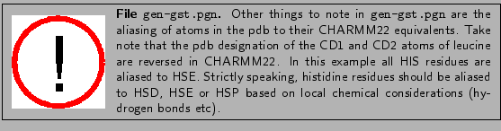
Now, to create a psf of the entire protein with glutathione chains bound, you will need to use the new topology file you created which has GGL and GLNK topology entries. The psfgen input file gen-gst.pgn has been provided for this. You should look at the file before using it. Notice how the GSH segment is handled:
| segment GSHA { | |
| first none | |
| pdb gsh-a.pdb | |
| } | |
| patch GLNK GSHA:1 GSHA:2 |
The command first none means that we apply no N-terminal or C-terminal generating patches to the GGL residue. The patch command is the means by which we apply our patch. It is followed by the name of the patch from the topology file (GLNK) and a list of residues and their segment names [segment:residue] to which the patch should be applied (GSHA:1 GSHA:2).
| source gen-gst.pgn |
Check the output to make sure no obvious errors occurred. If successful, you will have created files gst.pdb and gst.psf.
| mol new gst.psf | |
| mol addfile gst.pdb |
Have the hydrogen atoms on GSHA and GSHB been built properly? Unusual geometry could indicate a problem with your topology file.
| cp ../1-examine/par_all27_prot_lipid.inp . |
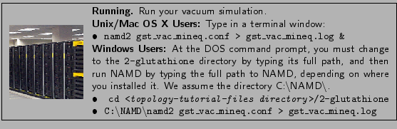
At this point, you are ready to run a simulation of your system in vacuum. If you have limited computational power (i.e. those performing this tutorial on a laptop), you should perform a vacuum run at this point and skip Section 2.3. If you have access to greater computational power (i.e. a small cluster), you may skip the vacuum run and continue on to Section 2.3.
| package require solvate | |
| solvate gst.psf gst.pdb -t 5 -o gst_solv |
| package require autoionize | |
| autoionize -psf gst_solv.psf -pdb gst_solv.pdb -is 0.2 -o gst_solv_ion |

As frames are added to the output trajectory (dcd) files, you can load them into VMD as a preliminary check that all is well with the topology file you created. Does the GSH molecule behave, or does the geometry distort? Unusual behaviour could indicate a problem in the topology file. If all appears well, you have succeeded!