tbss> vmd da.psf da_smd.dcd
|
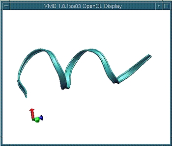
Position the molecule (rotate, translate, scale) so that you have the
view of the entire length of the molecule. Change the representation: Select the menu item Graphics -> Representations. In the Graphical Representations window, set Drawing Method on Ribbons. The Ribbon representation is good for viewing the overall structure, but you may explore any other representations or even multiple views. Use the scroll bar at the bottom of the VMD Main window to browse through the trajectory.
|  |
An important structural change during the helix-coil transition is the breaking of hydrogen bonds. You can monitor hydrogen bonds using VMD.
Choose CPK from Drawing Methods. This will change the representation of the molecule from Ribbon to CPK.
Now let's show hydrogen bonds:
| 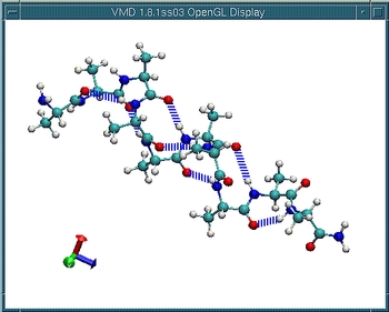 |
Again using the scroll bar at the bottom of the VMD Main window, browse through the trajectory. Observe the hydrogen bond breaking as the molecule is stretched.
Once you are done, quit VMD (the menu item File -> Quit).