You have probably obtained VMD in order to visualize molecules, so we'll load up one of the provided molecular structures to demonstrate the capabilities of VMD. On the button bar, click on the Mol button. This brings up the Molecules form. (As before, this form may be turned off by clicking the button in the center top of the form, labeled Molecules). Select Load From Files on the Molecules form to bring up the Files form.
We will load a PDB (Protein Data Bank) file containing the coordinates of the atoms in myoglobin (compliments of Joel Berendzen from the Biophysics Group at Los Alamos National Laboratory). In the browser on the left, select the line that says pdb only then click on the button labeled Select pdb. Use the file browser that appears to go to the subdirectory proteins/ of the VMD distribution (you may have to ask where this is located; try /usr/local/lib/vmd or /usr/local/vmd). Once there, select the file mbco.pdb. (Be careful when changing directories as the Forms library does not understand double clicks, and clicking too rapidly may cause the form to get very upset and core dump. This problem will be fixed in future versions of VMD.) Once the PDB file is selected, click on the Load Molecule button in the center of the Files form. You have loaded a myoglobin structure. Figure 2.1 shows an example of VMD displaying this protein.
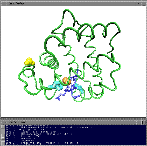
Figure 2.1: Sample VMD session displaying myoglobin.
You can use the mouse to manipulate the structure in the display window. There are three basic mouse modes: rotation, translation, scaling. The mode can be changed from the display window popup menu by pressing the right mouse button and picking the option under `Mouse Mode', or by pressing r, t, or s on the keyboard. Experiment with these modes, and note how the cursor changes to indicate the current mode. In rotation mode, the left mouse button controls rotation about axes parallel to the screen, and the middle button controls rotation about the axis perpendicular to the screen. In translation mode, the left mouse button controls translation parallel to the screen, while the middle button controls translation in and out of the screen. Finally, in scaling mode, both the left and middle buttons control global scaling when the mouse is moved left or right, but the middle button causes larger changes. In all modes, the right mouse button controls the display window popup menu.
By default the myoglobin bonds are represented as lines and non-bonded atoms as points, with the color in both cases representing the atom type. This representation is easy for the computer to draw but is not always informative, especially when there are a large number of atoms. VMD allows you to display many of the common molecular representations. To access these, open the Graphics form using the button bar.
Suppose you would like to view the myoglobin structure with its
protein backbone represented as a tube, the heme represented as
licorice, the ![]() ion and CO molecule represented as van der
Waals spheres, and histidines 64 and 93 represented as CPK
models. First, type backbone in the atom selection
text entry
area and press 'enter' to select the myoglobin backbone. All of the
protein except for the backbone will disappear. Choose drawing method
`Tube' from the drawing method chooser to render the backbone as a
tube, and chose coloring method `Backbone'
from the coloring method
chooser to color the tube with the predefined backbone color. Click
on the Create New button. This causes a new line to appear on
the browser identical to the first line. The new line can be changed
without affecting the first one, so clear the atom selection text area
and then enter resname HEM to select the heme. At this point
the heme isn't visible because it cannot be drawn as a tube, so choose
the `Licorice' drawing method
to make it appear. Click on Create
New again to make a new view, and enter resname SO4 CO to
select the
ion and CO molecule represented as van der
Waals spheres, and histidines 64 and 93 represented as CPK
models. First, type backbone in the atom selection
text entry
area and press 'enter' to select the myoglobin backbone. All of the
protein except for the backbone will disappear. Choose drawing method
`Tube' from the drawing method chooser to render the backbone as a
tube, and chose coloring method `Backbone'
from the coloring method
chooser to color the tube with the predefined backbone color. Click
on the Create New button. This causes a new line to appear on
the browser identical to the first line. The new line can be changed
without affecting the first one, so clear the atom selection text area
and then enter resname HEM to select the heme. At this point
the heme isn't visible because it cannot be drawn as a tube, so choose
the `Licorice' drawing method
to make it appear. Click on Create
New again to make a new view, and enter resname SO4 CO to
select the ![]() ion and the CO molecule, and choose the drawing
method `VDW' to render them as Van der Waal spheres. Once again,
press the Create New button and enter resid 93 64 to
select the two histidines, and render them as `CPK'. If you followed
all that, then congratulations, you have made a beautiful image of
myoglobin! Many more ways to represent atoms are possible. Please
experiment with the options available in the Graphics form.
ion and the CO molecule, and choose the drawing
method `VDW' to render them as Van der Waal spheres. Once again,
press the Create New button and enter resid 93 64 to
select the two histidines, and render them as `CPK'. If you followed
all that, then congratulations, you have made a beautiful image of
myoglobin! Many more ways to represent atoms are possible. Please
experiment with the options available in the Graphics form.